Blood Vessel Development
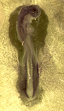
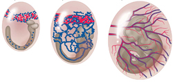 We all know that blood flow is crucial to keep us alive, but few people realize that blood flow also determined how we developed in the first place. The blood vessels of the embryo are initially established by a process called vasculogenesis which occurs in the absence of blood flow. These early vessels form a honey-combed shaped network known as the capillary plexus. With the onset of flow, these vessels are remodeled into more mature vasculature, typified by hierarchical branching morphology, changes in endothelial cell alignment and recruitment of peripheral cell types. Remodeling is dependent on blood flow and does not occur if blood flow is stopped. The regulation of circulatory development is critical to embryonic health. If the blood does not flow properly, the embryo does not develop and dies.
In adults, we know that the force blood exerts against a blood vessel wall determines how blood vessels branch and develop throughout the body. This force is called wall shear stress. We have recently showed that shear stresses are necessary for vascular remodelling. We are currently investigating whether other forces created by flowing blood, such as circumferential stretch, are also necessary, as well at looking if physical forces play a role in the initial vessel patterning.
Effect of Shear Stress on Angiogenesis
The first aim of my research is to understand the effects of wall shear stress on vascular remodelling in vivo. Vascular remodelling involves both the enlargement of some vessels, a process called arteriogenesis, and the formation of new blood vessels, termed angiogenesis. We have developed tools to visualize both flow dynamics and changes in vascular morphology in real-time using time-lapse microscopy. We use the embryonic vasculature as our model since it is easily accessible for imaging and undergoes drastic changes in a short period of time (24h).
Using high speed microscopy, we can image and calculate forces within the developing embryo. By physical and genetic manipulation, we can also alter these forces and see how this affects embryonic development. By time-lapse microscopy, we can watch these events occur in real time. Combining this with techniques from molecular biology, we can begin to understand the interplay of genetics and mechanics.
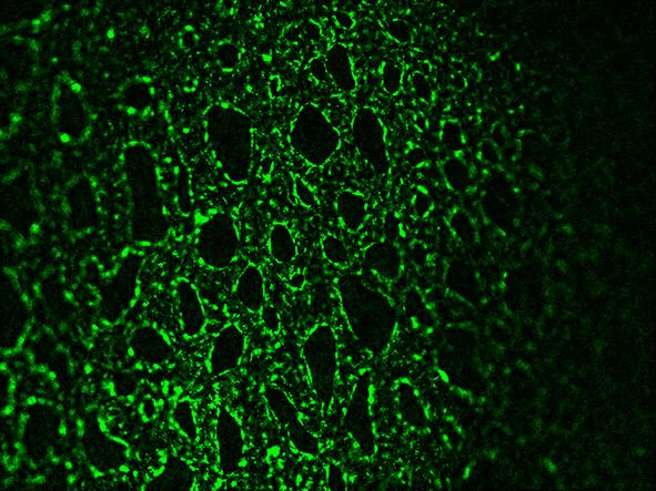
Time-Lapse of Vascular Remodelling in the Capillary Plexus. Red arrow shows vascular sprouting (angiogenesis). The large vessel on the left hand side forms by arteriogenesis (expansion of an existing vessel).
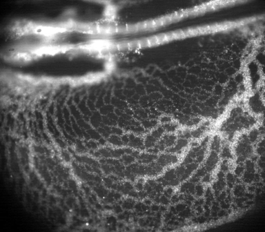
Blood Flow Dynamics During Vascular Development. We seed embryonic blood flow with small fluorescent microsphere and then image at high-speeds (250 fps) to calculate flow dynamics in the embryonic blood vessels. Published in Jones, E.A. (2011) “The Initiation of Blood Flow and Flow Induced Events in Early Vascular Development” Semin Cell Dev Biol, 22(9):1028-35. Abstract.
Ontogeny of Mechanotransduction
The second aim of my research is to study the development of shear-sensitive mechanisms during embryonic vascular development. Endothelial cells have the capacity to sense hemodynamic forces through the process of mechanotransduction. When blood flow starts in the embryo, many of the components required for sensing hemodynamic forces in the adult have not yet formed.
Adjunct Professor Department of Chemical Engineering, McGill
Current Position
Department of Cardiovascular Science
KU Leuven
3000 Leuven
Belgium