Image Gallery
Imaging of Antibody Staining
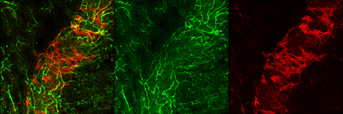
Alpha-smooth muscle actin staining of smooth muscle cells (red) around a blood vessel (green).
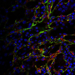
Staining of blood vessels for PECAM-1 (green), VEGFR2 (red) and Topro (blue).
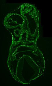
Collagen iV Staining on sections of an E9.5 mouse embryo
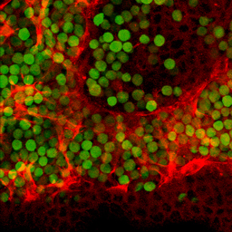
Anti-Alpha smooth muscle actin highlights smooth muscle cells (red) and green fluorescent protein (GFP, green) shows red blood cells.
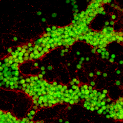
Antibody staining to VEGFR2 (red) showing GFP-expressing RBCs (green)
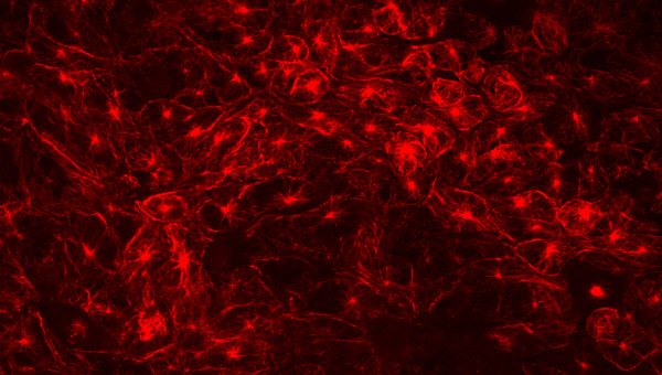
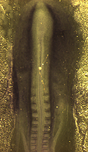
alpha-Tubulin Staining on Embryos
Adjunct Professor Department of Chemical Engineering, McGill
Current Position
Department of Cardiovascular Science
KU Leuven
3000 Leuven
Belgium